Follicular cysts (known as epidermal inclusion cysts or epidermoid cysts; sometimes erroneously referred to as sebaceous cysts) are a cystic dilatation of the upper portion of the outer sheath of the hair follicle lined by a layer of stratified cornifying epithelial cells indistinguishable from the epidermis.[1] These cysts may be solitary or multiple, firm to fluctuant, round and well-circumscribed.[2] Follicular cysts vary in size from 2 mm to > 5 cm.[1] They are found in about a third of the nonneoplastic, noninflammatory tumor-like lesions removed in dogs.[3] Predilection in middleaged to older dogs has been reported.[2]
Cysts may rupture releasing yellowish, brownish, or greyish material (keratin) into the surrounding dermis and subcutis. Keratin is highly irritating, evoking a strong inflammatory response and secondary bacterial infection.[4] The inflammation, especially with multiple cysts, may irritate the dog, lesions may become painful, pruritic, or both, causing the dog to constantly lick and chew the affected area, which exacerbates the problem.
The therapy of choice for multiple follicular cysts is surgical removal. Using the laser allows for minimal tissue removal so the wound edges can usually be sutured, whereas with the traditional scalpel more tissue will need to be excised and wound closure over this pressure point will be more troublesome. The laser is used at high power in the initial phase because at this point we are ablating tissue that consists mostly of keratin, which is low in water content so more power density is needed. Once the keratin is removed lower power is used which allows the surgeon to avoid coagulation of the normal underlying tissues. Post-op with the CO2 laser is less painful and has less swelling. And in areas where there aren’t large amounts of adjacent mobile skin to close the wound, like the elbow, less swelling results in superior postoperative tissue healing.

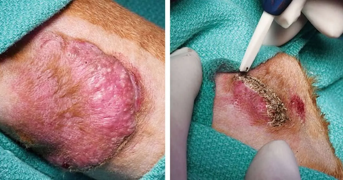
- Figure 1: Preoperative aspect of the left and right elbows with multiple follicular cysts.

The patient
Sadie, a 7.5-year-old female spayed Chesapeake Bay retriever, was brought with follicular cysts on both elbows (Figure 1). The cysts periodically had become inflamed and then ruptured with discharge. Initially, the problem improved temporarily with the use of Differin gel (retinol) and Mupirocin prescribed by the local veterinarian. However, by the time of the visit to our dermatology clinic these medications had stopped working, the elbows became extremely pruritic, and the dog constantly licked them. The problem with follicular cysts had persisted for one-and-a-half to two years and the symptoms were not seasonal. The dog had allergies and had been treated by her local veterinarian with SLIT allergen solution and a hypoallergenic diet.
Physical examination revealed moderately raised erythematous elbow callus areas. The right side had a long extended erythematous raised area with several hemorrhagic draining tracts. The left side callus was smaller but with similar hemorrhagic tracts. There were visible comedones with keratin easily expressed from both of these areas.
It was decided to ablate the follicular cysts from both elbow areas with a CO2 laser.
Anesthesia and pain management
The patient was premedicated with dexmedetomidine (DexDormitor) and butorphanol (Torbutrol) intravenously and received an injection of robenacoxib (Onsior).
General anesthesia was mask induced with isoflurane in oxygen and maintained via an endotracheal tube.
Laser equipment
VetScalpel CO2 laser model VS-4530 (by Aesculight, Bothell, Wash.) was utilized with a straight handpiece and a ceramic wide ablation tip (the tip is shown in Figures 2-A, 2-B, 2-C, and 3-A).
Laser settings
Power output: 30 W
- Laser mode: SuperPulse repeat F2-5 (21.75 W average power at 72.5 percent duty cycle)
- Pulse frequency: 29 Hz
- Pulse duration: 25 msec
- Focal laser spot size: 2.5 mm x 0.4 mm (area of approximately 1 mm2) For the ablation of deeper areas, the power and pulse duration were decreased and the settings were changed to the following: Power output: 15 W
- Laser mode: repeat non-SuperPulse F1-4 (6 W average power at 40 percent duty cycle)
- Pulse frequency: 20 Hz
- Pulse duration: 25 msec
- Focal laser spot size: 2.5 mm x 0.4 mm (area of approximately 1 mm2)

- Figure 2-A: Intra-op view. The larger section of the elbow skin with follicular cysts on the right elbow was first outlined with a single pass of the laser.

- Figure 2-B: Intra-op view. The edge of the lesion was grasped with a tissue forceps and retracted. The lesion was undermined with the laser beam and excised.

- Figure 2-C: Intra-op view. The remaining section of the elbow skin with multiple follicular cysts was ablated with several laser passes.

- Figure 2-D: Intra-op view. After each pass the surgical site was wiped with sterile saline on gauze pads until the surgeon ensured that no more cysts were left.
Surgical procedure
The surgical area was aseptically prepared, including the clipping of overlying hair. The larger section of the skin with follicular cysts on the right elbow was excised—it was first outlined with a single pass of the laser (Figure 2-A). Then, the edge of the lesion was grasped with a tissue forceps and retracted, while the lesion was undermined with the laser beam and excised (Figure 2-B). In the process, two larger blood vessels were severed and had to be ligated. The remaining section of the elbow skin with multiple follicular cysts was ablated with several laser passes (Figure 2-C). After each pass, the surgical site was wiped with sterile saline on gauze pads and gentle pressure was applied to express the contents of the remaining deeper follicular cysts until the surgeon ensured that no more cysts were left (Figure 2-D).
The affected area on the left elbow was much smaller. The follicular cysts were ablated in multiple laser passes (Figure 3-A), with the contents of the cysts expressed and wiped away between each pass (Figure 3-B). The laser procedure continued until no content could be expressed. All ablated tissue and debris were removed with a sterile gauze pad prior to wound closure (Figure 3-C).

- Figure 3-A: Intra-op view. The follicular cysts on the left elbow were ablated in multiple laser passes.

- Figure 3-B: The contents of the cysts were gently expressed and wiped away between each pass.

- Figure 3-C: All ablated tissue and debris were removed with a sterile gauze pad prior to wound closure.
Wound closure
For each elbow, surgical margins were apposed and sutured (Vicryl) was used for subcutaneous and nylon for cutaneous closure) (Figure 4). A soft wrap was then applied to help in holding the surgical margins together, as well as to decrease the swelling and to absorb any serum leakage. Then the suture site was treated with mupirocin 2% ointment and covered with a Telfa pad, cast padding and the usual outer bandage material. The patient was used to the DogLeggs, so this was a good way to help prevent her from bothering the bandage.

- Figure 4: Immediately post-op aspect of the surgical areas on left and right elbows, with sutures in place.

- Figure 5: Healed surgical areas 12 weeks post-op.
Postoperative care
To prevent infection at the site, the patient was put on a three-week course of oral cephalexin (500 mg capsules, two capsules b.i.d.). This was warranted because of all of the follicular contents coming out onto the surgical area.
For pain management, the patient was prescribed tramadol (50 mg tablets, 1.5 to 3 tablets once or twice daily, as needed).
Follow up
The patient was seen by a local veterinarian for bandage change at three days after the surgery, and then four days later. The bandages were changed in seven days. At the bandage changes, the suture sites were gently cleaned with chlorhexidine scrub and the area was patted dry. Then the suture site was covered with mupirocin 2% ointment, a Telfa pad, cast padding, and the usual outer bandage material and protected with the DogLeggs. Bandage change was continued every seven days until suture removal at three weeks post-op.
During the first week, there is typically some inflammation and serum seepage, which subside by the second week. Sutures are removed three weeks postoperatively.
Summary
This article describes a CO2 laser surgical procedure that in most cases proves curative multiple follicular cysts on canine elbows, such as swelling, inflammation, draining hemorrhagic tracts, pruritus, pain, and secondary infection. This laser procedure combined both excision and vaporization of the affected skin containing follicular cysts. Laser surgery allowed ablation of multiple layers of cysts and adjacent hair follicles without excessive thermal damage to the surrounding normal tissues.[5] The procedure was facilitated by the ability of the CO2 laser to coagulate small blood vessels during the surgery. The owner was happy with the outcomes and reported that the dog had recovered very well, with no concerns.
About the Author
David D. Duclos, DVM, DACVD, is a small-animal practitioner in Lynnwood, Wash., where he is the owner and clinical dermatologist at the Animal Skin & Allergy Clinic. He is an associate clinical instructor for the Western University College of Veterinary Medicine in Pomona, Calif., and teaches senior veterinary students as externs at his clinic. He frequently hosts veterinary students from other veterinary medical colleges around the U.S. and from Europe who are seeking to learn about the specialty of veterinary dermatology during their third and fourth year of veterinary school. In addition, he teaches veterinary residents in dermatology who are seeking to learn more about laser surgery for two- to four-week externships sponsored by the American College of Veterinary Dermatology. He has authored a number of book chapters and scientific papers on various subjects in veterinary dermatology, and lectures extensively in North America and Europe. Dr. Duclos is well known in the veterinary dermatology specialty for his expertise in CO2 laser surgery and for his interest in clinical photography.
References
- Villalobos AE. Epidermal and Hair Follicle Tumors. In: Aiello SE, Moses MA. Merck and the Immediately post-op aspect of the surgical areas on left and right elbows, with sutures in place. Healed surgical areas 12 weeks post-op. Figure 3-A: Intra-op view. The follicular cysts on the left elbow were ablated in multiple laser passes. Figure 3-B: The contents of the cysts were gently expressed and wiped away between each pass. Figure 3-C: All ablated tissue and debris were removed with a sterile gauze pad prior to wound closure. Merck Veterinary Manual. 11th ed, https://www.merckvetmanual.com/integumentary-system/tumors-of-the-skin-and-soft-tissues/epidermal-and-hair-follicle-tumors. Accessed 2-7-18.
- Raskin RE. Skin and subcutaneous tissues. In: Raskin RE, Meyer D. Canine and feline cytology: a color atlas and interpretation guide. 3rd ed, St. Louis, MO: Saunders, 2010;34-90.
- Raskin RE. Advanced Cytology on Skin Mass. Paper presented at: International Congress of the Italian Association of Companion Animal Veterinarians; May 19-21 2006; Rimini Italy. http://www.ivis.org/proceedings scivac/2006/raskin6_en.pdf. Accessed 2-15-18.
- Tvedten H. Cytology of inflammatory or neoplastic masses. In: Willard MD, Tvedten H. Small animal clinical diagnostics by laboratory methods. 5th ed, St. Louis, MO: Elseveir/Mosby, 2012;337-363.
- Wilder-Smith P, Arrastia AM, Liaw LH, Berns M. Incision properties and thermal effects of three CO2 lasers in soft tissue. Oral Surg Oral Med Oral Pathol Oral Radiol Endod. 1995;79(6):685-91.