Draining an infected epidermal cyst on the ear lobule involves a minor surgical procedure that must be done under sterile conditions by a healthcare professional. Below is a detailed, step-by-step clinical outline of how this is typically done.
🦻 Drainage of Infected Epidermal Cyst – Ear Lobule
⚠️ Important
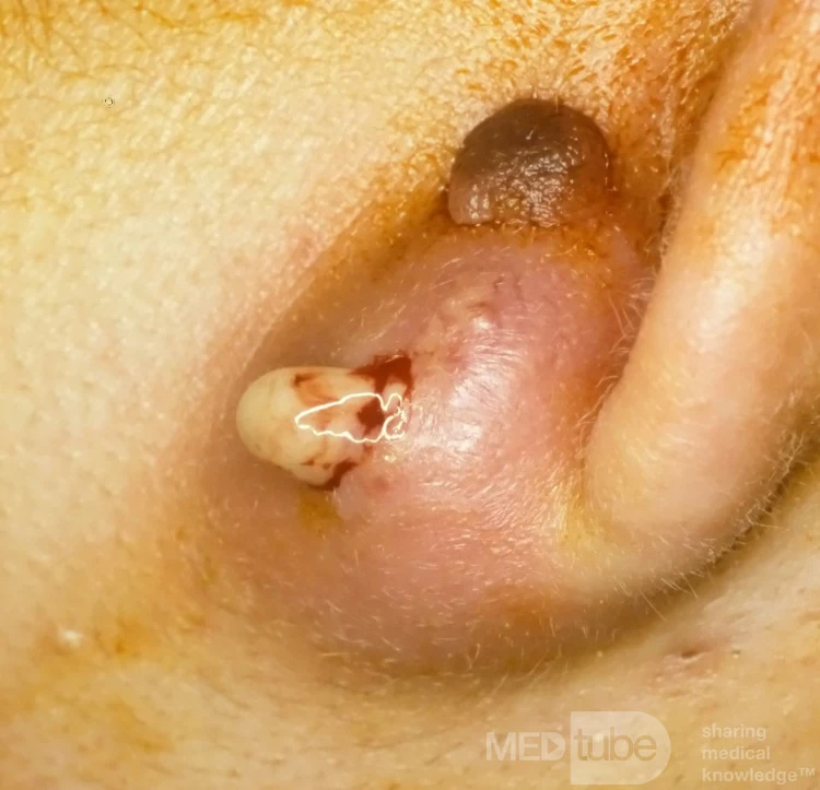
If the cyst is infected, the area is usually red, swollen, tender, and possibly draining pus. In this case, complete excision is not done immediately; drainage is performed first, followed by excision later after inflammation subsides.
🛠️ Materials Needed
-
Sterile gloves
-
Local anesthetic (e.g. lidocaine 1% with epinephrine)
-
Antiseptic (e.g. povidone-iodine or chlorhexidine)
-
11 or 15-blade scalpel
-
Sterile gauze
-
Hemostats or forceps
-
Dressing materials
-
Syringe and needle (for anesthesia)
-
Optional: Culture swab for pus
🔬 Step-by-Step Procedure
1. Preparation
-
Position the patient comfortably.
-
Clean the ear lobule and surrounding skin with antiseptic.
-
Drape the area for a sterile field.
2. Anesthesia
-
Inject local anesthetic around the cyst (not directly into it to avoid spreading the infection).
3. Incision and Drainage
-
Use a scalpel to make a small linear incision directly over the most fluctuant (softest, pus-filled) part of the cyst.
-
Allow the purulent (pus) material to drain freely.
-
Gently squeeze around the cyst to evacuate contents. Use sterile gauze to soak excess drainage.
4. Cavity Cleaning
-
DO NOT try to remove the cyst wall (sac) during infection—it increases the risk of spreading infection and bleeding.
-
Irrigate the cavity with sterile saline to flush debris.
5. Packing (optional)
-
If there’s a large cavity, insert a small piece of iodoform gauze to keep the incision open and allow further drainage.
-
Packing may be changed daily until the infection resolves.
6. Dressing
-
Apply a sterile dressing over the incision site.
💊 Post-Procedure Care
-
Antibiotics (e.g. amoxicillin-clavulanate or doxycycline) may be prescribed, especially if cellulitis is present.
-
Pain management: Acetaminophen or ibuprofen.
-
Follow-up: Recheck in 2–3 days. Once infection resolves (usually in 1–2 weeks), complete surgical excision of the cyst sac can be done to prevent recurrence.
📌 Important Notes
-
Never excise an infected cyst—wait until it has healed.
-
If recurrent, full cyst removal should be scheduled after inflammation resolves.
-
Ear lobule cysts can cause cosmetic concerns, so excision should be meticulous.
🔗 References
🦻 Infected Epidermoid Cyst Drainage – Ear Lobule
⚠️ Important Note
If the cyst is infected, immediate excision is not recommended due to increased risk of complications. The initial approach involves drainage to alleviate symptoms and reduce infection. Once inflammation subsides, definitive surgical removal can be performed.
🛠️ Materials Needed
-
Sterile gloves
-
Local anesthetic (e.g., 1% lidocaine with epinephrine)
-
Antiseptic solution (e.g., povidone-iodine or chlorhexidine)
-
11 or 15 blade scalpel
-
Sterile gauze pads
-
Hemostats or forceps
-
Sterile saline solution
-
Sterile dressing materials
-
Optional: Culture swab for pus
🔬 Step-by-Step Procedure
1. Preparation
-
Positioning: Ensure the patient is seated comfortably with adequate lighting.
-
Sterilization: Cleanse the surrounding skin with an antiseptic solution.
-
Draping: Place sterile drapes around the affected area to maintain a sterile field.
2. Anesthesia
-
Injection: Administer local anesthesia around the cyst using a 1% lidocaine solution.
-
Onset: Allow approximately 5–10 minutes for the anesthetic to take effect.
3. Incision and Drainage
-
Incision: Make a small linear incision over the most fluctuant (softest) part of the cyst using an 11 or 15 blade scalpel.
-
Drainage: Gently express the cyst to evacuate the purulent material.
-
Irrigation: Flush the cavity with sterile saline to remove debris and reduce bacterial load.
4. Cavity Management
-
Debridement: If necessary, use sterile forceps or curettes to remove any necrotic tissue.
-
Packing: In some cases, a small piece of sterile gauze may be placed inside the cavity to promote drainage and prevent premature closure.
5. Dressing
-
Application: Apply a sterile dressing to the incision site.
-
Instructions: Instruct the patient to keep the area clean and dry, and to change the dressing as recommended.
💊 Post-Procedure Care
-
Antibiotics: Prescribe oral antibiotics if there is evidence of surrounding cellulitis or systemic infection.
-
Pain Management: Recommend over-the-counter analgesics such as acetaminophen or ibuprofen for pain relief.
-
Follow-Up: Schedule a follow-up appointment in 1–2 weeks to assess healing and discuss definitive surgical removal if necessary.
📌 Important Considerations
-
Avoid Immediate Excision: Performing excision during active infection can increase the risk of complications and recurrence.
-
Sterility: Maintain strict aseptic technique throughout the procedure to prevent further infection.
-
Documentation: Document the procedure details, including the amount and appearance of the drained material, and any complications encountered.
🔗 References
-
Mayo Clinic: Epidermoid Cysts – Diagnosis and Treatment
-
Children’s Healthcare of Atlanta: Epidermoid Cyst, Infected (Incision and Drainage)
-
StatPearls: Epidermal Inclusion Cyst – StatPearls – NCBI Bookshelf
1. ⚠️ If the cyst is infected (red, painful, swollen, possibly draining pus):
✅ Step 1: Incision and Drainage (I&D)
-
Performed by: A medical professional (dermatologist, GP, or ENT surgeon)
-
What happens:
-
Local anesthesia is given.
-
A small cut is made to drain pus and reduce pressure.
-
The cavity may be flushed with sterile saline.
-
In some cases, gauze or a drain is placed to keep the wound open for ongoing drainage.
-
✅ Step 2: Antibiotic Therapy
-
If there is surrounding cellulitis (skin infection) or risk of spreading:
-
Oral antibiotics such as:
-
Amoxicillin-clavulanate (e.g., Augmentin)
-
Clindamycin (if allergic to penicillin)
-
Doxycycline (covers MRSA in some regions)
-
-
Duration: 5–10 days, depending on severity
-
✅ Step 3: Wound Care
-
Daily gentle cleaning with saline
-
Keep the area dry and protected
-
Avoid squeezing or manipulating the area
-
Follow-up to assess healing and remove any packing
2. 🧘♀️ After the infection resolves (usually in 1–3 weeks):
✅ Step 4: Complete Surgical Excision
-
Why: To prevent recurrence, the entire cyst wall or sac must be removed.
-
Procedure:
-
Done under local anesthesia
-
A small incision is made along natural skin lines
-
Cyst sac is dissected and removed in full
-
Sutures may be used to close the wound
-
Minimal scarring if done properly
-
❌ Do NOT do this:
-
Do not squeeze or pop the cyst at home.
-
Do not apply over-the-counter “boil creams” to an actively infected cyst.
-
Do not attempt home removal—it can worsen infection or cause scarring.
🔁 Summary Flowchart
| Cyst Condition | Treatment |
|---|---|
| Not infected | Elective excision (complete removal) |
| Infected, swollen | I&D + antibiotics |
| Drained, healing | Daily wound care + optional antibiotics |
| After infection heals | Definitive excision to prevent recurrence |
📚 Clinical References
-
Mayo Clinic: Epidermoid Cyst Treatment
-
StatPearls (NIH):Epidermal Inclusion Cyst
-
DermNet NZ: Epidermoid Cyst Management