Dr. Blackhead Extractions EP:251 – REMOVAL PIMPLE

What causes a sebaceous cyst?  

Sebaceous cysts are formed within the sebaceous gland, which is the gland which produces sebum. These cysts develop when the hair follicles become clogged due to a build up of sebum or keratin. These cysts can also be formed from pimples or as a result of trauma to the sebaceous glands. Individuals with a genetic predisposition such as steatocystoma multiplex, Gardner’s syndrome or Basal Cell Nevus Syndrome are also prone to developing sebaceous cysts.

How do you diagnose a sebaceous cyst?

A diagnosis of a sebaceous cyst can be determined by a physical examination of the nodule by a dermatologist, family physician or other healthcare provider. There are occasions when additional testing is required to make a definitive diagnosis of a cyst, since it can sometimes be mistaken for a different type of skin tumor.

Common tests used to diagnosis a sebaceous cyst include:

  • Cat scan – This test is performed to rule out other abnormalities or cancer.
  • Ultrasound – This test is performed to establish the contents of the cyst and depth of inflammation.
  • Punch biopsy – This test is performed to identify the histology of the cyst.
  • Culture and Sensitivity – This exam is performed to determine the type of bacteria responsible for the infection and the best antibiotic to treat the infection.

  • Physical Exam: The provider will check for a lump or swelling under the skin. A sebaceous cyst usually feels firm, round, and mobile beneath the skin. It may be located anywhere on the body but is commonly found on the face, neck, or back.
  • History: The provider may ask about the symptoms, like when the lump first appeared, if it has grown in size, or if it’s causing pain or discomfort. A sebaceous cyst is often painless unless it becomes infected or inflamed.
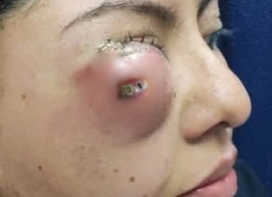
  • Visual Inspection: The cyst might have a small opening (a pore) in the center, through which the contents may drain if the cyst becomes infected or ruptures.
  • Possible Additional Tests: If there’s any doubt about the diagnosis, or if the cyst is large or causing complications, further tests might be recommended. This could include:
    • Ultrasound: To help determine the size and structure of the cyst.
    • Biopsy: In rare cases, a biopsy may be done to rule out other conditions, especially if the cyst appears unusual.
Certainly! Here’s a more detailed explanation of diagnosing a sebaceous cyst, including references from medical sources:

1. Clinical History and Physical Examination

The diagnosis of a sebaceous cyst generally begins with a thorough medical history and physical examination. A sebaceous cyst is often identified based on its characteristic features:

  • History: The patient is asked about the size, location, onset, and progression of the cyst. In many cases, the cyst has been present for a long time and grows slowly. A history of trauma or blockage of the sebaceous gland can be significant.
  • Physical Examination: The healthcare provider will feel the lump, typically finding a round, firm, and mobile mass beneath the skin. It is typically painless unless it becomes infected or inflamed.
    • Skin over the cyst: The skin may appear normal or slightly red or inflamed, particularly if the cyst is infected or ruptured.
    • Pore or opening: A central pore (opening) may be present, which is a characteristic feature of sebaceous cysts. This opening may expel a cheesy, foul-smelling substance if the cyst is ruptured or infected (keratin, sebum).

2. Differential Diagnosis

While sebaceous cysts are common, they can be confused with other skin conditions or masses. The differential diagnosis includes:

  • Epidermoid cysts: Similar to sebaceous cysts but arising from the epidermis.
  • Lipomas: Soft, rubbery, and usually mobile benign fatty tumors, distinct from sebaceous cysts in texture.
  • Abscesses: Infected cysts may present similarly to abscesses but typically have a central opening or a smooth, firm texture.
  • Dermoid cysts: These can be deeper and may contain hair, skin, and sebaceous material.

3. Additional Diagnostic Tests

In most cases, the diagnosis of a sebaceous cyst is made clinically, but additional tests may be required if the cyst presents with unusual features or complications (e.g., infection or rapid growth).

Ultrasound:

Ultrasound imaging is often used when the cyst is deep or difficult to assess. It can help confirm the cyst’s nature, size, and location. Sebaceous cysts typically appear as well-defined, cystic structures with a hypoechoic (dark) appearance due to the presence of keratin or sebum inside.

Reference:

  • Ormenese et al., “Use of ultrasonography for the diagnosis and follow-up of epidermal cysts” Dermatology, 2009.

Fine Needle Aspiration (FNA):

In cases where the diagnosis is uncertain or to rule out malignancy, fine needle aspiration may be performed to extract a sample from the cyst. This is usually done when there is suspicion of a different underlying condition.

Biopsy:

A biopsy is generally not needed for typical sebaceous cysts, but it might be recommended if there’s concern about a more serious condition, such as skin cancer or a tumor that mimics a cyst.

Reference:

  • Madan, V., et al., “Atypical presentation of sebaceous cysts: a review of cases in a dermatology practice” Clinical and Experimental Dermatology, 2012.

4. Infection and Inflammation

Sebaceous cysts can become infected or inflamed, causing changes that might prompt medical intervention. Infected cysts may become red, swollen, and painful, and the overlying skin may become warm to the touch. In these cases, drainage, antibiotics, or even surgical removal might be required.

Infected Cysts:

An infected sebaceous cyst can present with symptoms like fever, increased redness, and pus drainage. If the cyst ruptures, the material inside can spill into the surrounding tissue, potentially causing an abscess.

Reference:

  • Patel, A., et al., “Infected sebaceous cysts: pathophysiology, diagnosis, and management” Journal of Clinical and Aesthetic Dermatology, 2018.

5. Management and Treatment

  • Observation: If asymptomatic, sebaceous cysts may simply be monitored for changes.
  • Surgical Excision: If the cyst becomes bothersome, infected, or grows large, surgical removal is typically recommended. This procedure involves excising the cyst completely, including its capsule, to prevent recurrence. Reference:
    • Thompson, S. et al., “Management of epidermoid cysts” The Journal of Dermatology, 2015.

Conclusion

A sebaceous cyst is usually diagnosed through clinical history and physical examination, with ultrasound or fine needle aspiration used for confirmation in certain cases. While most sebaceous cysts are benign, monitoring for signs of infection or malignancy is essential. For typical cases, treatment may involve observation or surgical removal if necessary.

By Davan

Leave a Reply

Your email address will not be published. Required fields are marked *