Scroll Down to watch👇👇
What causes a sebaceous cyst?
Sebaceous cysts are formed within the sebaceous gland, which is the gland which produces sebum. These cysts develop when the hair follicles become clogged due to a build up of sebum or keratin. These cysts can also be formed from pimples or as a result of trauma to the sebaceous glands. Individuals with a genetic predisposition such as steatocystoma multiplex, Gardner’s syndrome or Basal Cell Nevus Syndrome are also prone to developing sebaceous cysts.
How do you diagnose a sebaceous cyst?
A diagnosis of a sebaceous cyst can be determined by a physical examination of the nodule by a dermatologist, family physician or other healthcare provider. There are occasions when additional testing is required to make a definitive diagnosis of a cyst, since it can sometimes be mistaken for a different type of skin tumor.
Common tests used to diagnosis a sebaceous cyst include:
- Cat scan – This test is performed to rule out other abnormalities or cancer.
- Ultrasound – This test is performed to establish the contents of the cyst and depth of inflammation.
- Punch biopsy – This test is performed to identify the histology of the cyst.
- Culture and Sensitivity – This exam is performed to determine the type of bacteria responsible for the infection and the best antibiotic to treat the infection.
Diagnosing a sebaceous cyst typically involves a physical examination and, in some cases, additional tests. Here’s how the process generally works:
- Medical History: The healthcare provider may start by asking about your medical history, including any previous skin conditions, the duration of the cyst, and whether there have been any changes in its size or appearance.
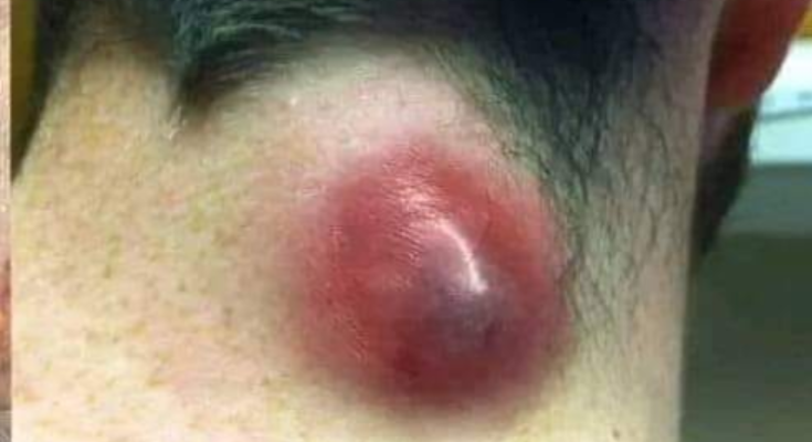
- Physical Examination: The doctor will examine the cyst. A sebaceous cyst usually appears as a small, round bump under the skin, typically on the face, neck, or back. It may have a central blackhead or small pore. The cyst is usually firm, mobile, and painless unless infected.
- Symptoms Assessment: The doctor will inquire if the cyst is causing discomfort, tenderness, or if it’s swollen or inflamed. If it is infected, it might be red, warm, and painful.
- Possible Draining or Culture: If the cyst is infected or there’s concern about other conditions, the doctor might drain the contents or take a sample (culture) to check for infection or other potential causes.
- Imaging (if needed): In rare cases, if the diagnosis is unclear or if there’s concern about other types of cysts or growths, imaging tests like an ultrasound or MRI may be used to assess the cyst’s structure and ensure it’s not something more serious, such as a lipoma or abscess.
- Biopsy (if necessary): If the doctor is uncertain about the diagnosis or if the cyst shows unusual features, they might recommend a biopsy to rule out other conditions, such as skin cancer.
In most cases, a sebaceous cyst can be diagnosed with a simple visual and physical exam. However, if the cyst appears unusual or causes complications, further diagnostic tests may be necessary.
1. Patient History
The first step in diagnosing a sebaceous cyst involves gathering relevant patient history. Key aspects to consider include:
- Duration: How long has the cyst been present?
- Changes in size: Has the cyst grown, remained the same, or decreased in size?
- Symptoms: Does the cyst cause pain, redness, or discharge?
- Previous Cyst History: Any history of similar cysts in the past?
- Trauma: Any recent injury to the skin or follicle where the cyst formed?
Reference: Fitzpatrick’s Dermatology in General Medicine (2019) notes that sebaceous cysts, also known as epidermoid cysts, often develop as a result of blockage or damage to the sebaceous gland, which leads to the accumulation of keratin and debris.
2. Physical Examination
During a physical exam, healthcare providers typically observe the following characteristics of sebaceous cysts:
- Appearance: Sebaceous cysts typically appear as a round, firm, movable bump under the skin, often with a visible central pore or blackhead at the surface.
- Location: Sebaceous cysts are most commonly found on the face, scalp, neck, or upper back.
- Size: They can vary in size, from small and barely noticeable to larger masses.
Reference: According to Dermatology: A Visual Guide by Jean L. Bolognia, sebaceous cysts are benign, non-inflammatory lesions filled with keratin, and they have a characteristic appearance that differentiates them from other dermatological conditions like lipomas or abscesses.
3. Assessing for Symptoms of Infection or Inflammation
- Infected cysts: If a sebaceous cyst becomes infected, it may become red, warm to the touch, tender, and possibly drain pus or foul-smelling material. In some cases, an abscess may form.
- Pain: Cysts that are inflamed or infected can be painful, while non-infected cysts are typically asymptomatic.
Reference: Mayo Clinic Proceedings (2007) emphasizes that infected sebaceous cysts may present with these symptoms and may require intervention such as drainage or antibiotics.
4. Differentiating from Other Skin Conditions
Several conditions can mimic sebaceous cysts, so it’s important to distinguish them from:
- Lipomas: Soft, fatty lumps that are typically mobile under the skin but lack the central pore characteristic of sebaceous cysts.
- Abscesses: Infected, painful lumps often accompanied by fever and systemic symptoms.
- Dermoid cysts: These may contain hair, skin, and teeth and are usually located near the eyes or scalp.
Reference: According to Dermatology: An Illustrated Colour Text by R. J. F. W. G. Marks, differentiation of sebaceous cysts from other cutaneous tumors is crucial to avoid misdiagnosis.
5. Possible Draining or Culture of Cyst Content
If the cyst is infected or if there is suspicion of another pathology, the doctor may drain the cyst and/or take a culture of the contents for further analysis. This can help identify bacteria or fungi, especially in cases of infection.
- Microscopic examination: If the cyst has been drained, the contents may be examined under a microscope to confirm its keratinous nature or to rule out infection.
- Culture: If there is an active infection, a culture can help identify the microorganism involved, aiding in antibiotic selection.
Reference: The Journal of Clinical and Aesthetic Dermatology (2016) discusses the importance of culturing cyst content in cases of infection to guide appropriate antibiotic therapy.
6. Imaging Tests (if necessary)
Imaging is rarely required for diagnosing a typical sebaceous cyst, but in certain cases, it may be used to:
- Confirm the diagnosis in the case of large or atypical cysts.
- Assess the depth or structure of the cyst if there is a concern about malignancy or another underlying pathology.
- Evaluate the surrounding tissues, especially if the cyst is near critical areas like nerves or blood vessels.
Ultrasound: A non-invasive imaging technique that may help visualize the contents and size of the cyst.
MRI: This may be used in rare cases where deeper or more complex cysts need to be evaluated, especially if there’s suspicion of a dermoid cyst or a malignancy.
Reference: American Family Physician (2016) mentions that imaging is generally not necessary unless the cyst is unusual or has complications like infection or deep-seated growth.
7. Biopsy (if necessary)
In very rare cases, if the sebaceous cyst appears unusual (e.g., irregular borders, rapid growth, or atypical features), a biopsy may be performed to rule out malignancy, such as basal cell carcinoma or squamous cell carcinoma. A biopsy involves removing a small sample of tissue from the cyst and examining it under a microscope.
Reference: According to the British Journal of Dermatology (2018), a biopsy is recommended if the clinical features suggest a possibility of malignancy or if there is significant doubt regarding the diagnosis.